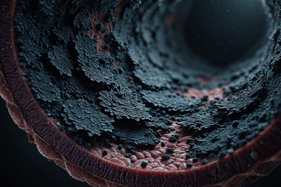
Endothelial lining.
CLINICAL PERSPECTIVE / NOVEL CONCEPT
Endothelial Wall Armoring:
Targeted Nanoparticle Aggregation to the Cerebral Microvasculature
as a Structural Neuroprotective Strategy Against Small Vessel Disease
Department of Neuroscience & Vascular Neurology
Translational Neuromodulation & Vascular Medicine Laboratory
Keywords: Endothelial Nanoparticles · Vascular Neuroprotection · Small Vessel Disease · ICAM-1 · VCAM-1 · eNOS · Glycocalyx · Cerebral Arteriole · Lipohyalinosis · Neurovascular Unit
June 2026
Ghost Publication Metadata
slug: endothelial-wall-armoring-nanoparticle-cerebral-svd
tags: Nanomedicine · Vascular Neuroprotection · Small Vessel Disease · Clinical Perspective · Dementia Prevention
excerpt (60 words): Cerebral Small Vessel Disease progresses through endothelial dysfunction, eNOS uncoupling, and lipohyalinosis of penetrating arterioles — mechanisms current pharmacotherapy cannot structurally reverse. This perspective introduces Endothelial Wall Armoring via targeted nanoparticle aggregation to the cerebral microvasculature as a novel structural neuroprotective strategy, anchoring bioactive nanocarriers to ICAM-1, VCAM-1, and glycocalyx-deficient endothelium to restore vascular homeostasis and arrest neurovascular lesion progression.
ABSTRACT
Abstract (60 words): Cerebral Small Vessel Disease progresses through endothelial dysfunction, eNOS uncoupling, and lipohyalinosis of penetrating arterioles — mechanisms current pharmacotherapy cannot structurally reverse. This perspective introduces Endothelial Wall Armoring via targeted nanoparticle aggregation to the cerebral microvasculature as a novel structural neuroprotective strategy, anchoring bioactive nanocarriers to ICAM-1, VCAM-1, and glycocalyx-deficient endothelium to restore vascular homeostasis and arrest neurovascular lesion progression.
Cerebral Small Vessel Disease (SVD) constitutes the principal pathological substrate of vascular cognitive impairment and contributes substantially to the pathophysiology of mixed dementia. Its progression is driven by the progressive structural deterioration of penetrating arterioles and cerebral capillaries, mediated by endothelial dysfunction, loss of glycocalyx integrity, eNOS uncoupling, oxidative stress, and lipohyalinosis of the vessel wall. Current pharmacological and lifestyle interventions address systemic risk factors but provide no mechanism for structural stabilisation of the already-compromised cerebral microvasculature. This perspective proposes a novel conceptual framework — Endothelial Wall Armoring (EWA) — based on the targeted aggregation of functionalised nanoparticles to the luminal surface of dysfunctional cerebral arteriolar endothelium, exploiting the upregulated expression of adhesion molecules (ICAM-1, VCAM-1, E-selectin) and the degraded glycocalyx as molecular anchoring platforms.
The proposed nanoparticle construct would serve not merely as a drug delivery vehicle — the dominant paradigm in cerebrovascular nanomedicine — but as a structural bioactive scaffold anchored to the endothelial surface, locally delivering: (i) eNOS activators to restore nitric oxide bioavailability and vasodilatory function; (ii) reactive oxygen species (ROS) scavengers to interrupt the oxidative cascade driving endothelial apoptosis; (iii) glycocalyx precursor components for luminal surface restoration; and (iv) anti-inflammatory mediators to suppress the VCAM-1/ICAM-1 inflammatory amplification loop. This approach reframes vascular neuroprotection from systemic pharmacological control to localised endothelial structural reinforcement — a paradigm shift with profound implications for the prevention of silent microinfarcts and neurovascular coupling failure in the pre-clinical phase of dementia.
1. INTRODUCTION: THE UNADDRESSED STRUCTURAL VULNERABILITY
1.1 The Endothelium as the Primary Lesion Site in SVD
Cerebral Small Vessel Disease encompasses a spectrum of pathological processes affecting the small perforating arteries (diameter 40–200 μm), arterioles, capillaries, and venules of the brain. Unlike large-vessel atherosclerotic disease, SVD operates through a microangiopathic mechanism in which the primary lesion site is the endothelial cell layer of the vessel wall itself. The endothelium of cerebral penetrating arterioles is subjected to unique haemodynamic stresses — including the high transmural pressure gradients of hypertension, the rheological alterations of diabetic microangiopathy, and the metabolic insults of dyslipidaemia — that cumulatively produce a dysfunctional endothelial phenotype characterised by increased permeability, inflammatory activation, and loss of vasomotor regulatory capacity.
The pathological sequence in SVD has been progressively elucidated: initial endothelial activation leads to upregulation of adhesion molecules (VCAM-1, ICAM-1, E-selectin), facilitating leucocyte adhesion and transmigration; progressive oxidative stress, mediated by NADPH oxidase activation and mitochondrial dysfunction, depletes nitric oxide bioavailability through eNOS uncoupling; the resulting loss of NO-dependent vasodilation and the deposition of plasma proteins through a disrupted blood-brain barrier (BBB) initiates lipohyalinosis — the pathological hallmark of advanced SVD, in which the smooth muscle layer of arterioles is replaced by acellular fibrous and hyaline material, permanently ablating the vessel's vasomotor responsiveness.
This progression from endothelial activation to lipohyalinosis represents a sequence of irreversible structural events for which no current therapeutic intervention provides direct structural counteraction at the vessel wall level. Systemic antihypertensives reduce haemodynamic stress but cannot reverse established lipohyalinosis; antiplatelet agents reduce thrombotic risk but do not restore endothelial function; statins reduce systemic inflammation but exert limited direct effect on the already-compromised arteriolar wall. The structural vulnerability of the cerebral microvasculature in SVD therefore represents a therapeutic gap — the precise gap that the Endothelial Wall Armoring concept is designed to address.
1.2 The Glycocalyx: A Neglected Therapeutic Target
The endothelial glycocalyx — a complex layer of glycoproteins, proteoglycans, and glycosaminoglycans covering the luminal surface of all vascular endothelial cells — plays a critical mechanosensory and protective role in vascular homeostasis. It acts as a mechanotransducer of fluid shear stress, a molecular sieve regulating macromolecular permeability, a reservoir for eNOS-activating signalling molecules, and a physical barrier preventing direct leucocyte-endothelium contact.
In SVD, the glycocalyx is systematically degraded by the very mechanisms that drive disease progression: oxidative stress cleaves heparan sulphate chains; hyperglycaemia impairs syndecan expression; hypertensive shear stress promotes shedding of glycocalyx components into the circulation. The resulting glycocalyx-denuded endothelial surface exhibits profoundly amplified inflammatory responsiveness, reduced NO production, and increased thrombogenicity. Critically, the degraded glycocalyx exposes underlying CD31 (PECAM-1) and CD44 epitopes — molecular targets that can be exploited for precision nanoparticle anchoring to dysfunctional, but not healthy, endothelium.
2. THE NANOPARTICLE CONSTRUCT: DESIGN PRINCIPLES FOR ENDOTHELIAL ANCHORING
2.1 Targeting Strategy: Exploiting Pathological Upregulation
The targeting strategy of the proposed EWA nanoparticle construct is predicated on a fundamental principle of precision nanomedicine: pathological tissue overexpresses molecular markers that healthy tissue does not, enabling selective localisation of functionalised nanocarriers to the disease site while sparing normal vasculature. In SVD, the dysfunctional endothelium of cerebral arterioles presents an array of upregulated surface molecules that are absent or minimally expressed on intact endothelium — constituting a molecular address code for precision targeting.
The principal targeting ligands proposed for the EWA construct are: (i) anti-ICAM-1 antibody fragments — ICAM-1 (Intercellular Adhesion Molecule-1) is systematically upregulated on endothelium subjected to inflammatory activation, oxidative stress, and hypertensive haemodynamic injury, providing a well-validated targeting epitope with extensive prior literature in vascular-targeted drug delivery; (ii) VCAM-1-binding peptides — VCAM-1 expression marks early endothelial activation and correlates directly with SVD lesion burden; (iii) anti-CD31 (PECAM-1) targeting moieties — CD31 is exposed on glycocalyx-denuded endothelium, providing a targeting opportunity specific to structurally compromised vessels; and (iv) E-selectin-binding peptides — particularly relevant in the context of inflammatory endothelial activation associated with SVD progression.
The multi-ligand targeting approach — combining two or more of these epitopes on a single nanoparticle construct — is expected to confer both targeting specificity and avidity sufficient to maintain firm nanoparticle adhesion under the pulsatile shear stress conditions of cerebral arterioles, a critical engineering parameter validated in prior in vitro microfluidic studies of vascular-targeted carriers.
2.2 Nanoparticle Architecture and Bioactive Cargo
The proposed nanoparticle architecture is based on a PLGA (poly-lactic-co-glycolic acid) polymeric core — a biodegradable, FDA-approved polymer with well-established safety profile, tunable degradation kinetics, and capacity for sustained release of encapsulated cargo — functionalised on its surface with the targeting ligands described above and coated with a polyethylene glycol (PEG) corona to extend circulatory half-life and reduce non-specific protein adsorption (opsonisation).
The bioactive cargo of the EWA construct is designed to address the four principal pathological drivers of endothelial deterioration in SVD simultaneously:
First, eNOS activators and NO donors — compounds including L-arginine, tetrahydrobiopterin (BH4) precursors, and inorganic nitrite — would be encapsulated to restore nitric oxide bioavailability at the endothelial surface, re-establishing NO-dependent vasodilation and anti-inflammatory signalling. A nanomaterial mimicking eNOS enzymatic activity has been described using MoS2 nanostructures that release NO while avoiding toxic peroxynitrite (ONOO⁻) production, demonstrating therapeutic effects in cerebrovascular ischaemia models.
Second, ROS scavengers — inorganic nanoparticle components including CeO2 (cerium oxide) and MnO2, which possess intrinsic superoxide dismutase and catalase-like enzymatic activity, would be incorporated to interrupt the oxidative cascade driving eNOS uncoupling, endothelial apoptosis, and glycocalyx degradation. Cerium oxide nanoparticles have demonstrated efficacy as free radical scavengers in cerebrovascular ischaemia models, with evidence of vascular repair promotion.
Third, glycocalyx precursor components — including heparan sulphate, hyaluronic acid, and syndecan precursors — would be incorporated to promote reconstitution of the degraded glycocalyx layer. Patent literature describes nanoparticle formulations carrying preassembled glycocalyx components targeted via anti-CD31 antibodies to endothelial surfaces, with demonstrated reconstitution of glycocalyx function in vitro.
Fourth, anti-inflammatory mediators — including rapamycin (an mTOR inhibitor with potent anti-inflammatory and antiproliferative effects on vascular smooth muscle) and short-interfering RNA (siRNA) sequences targeting NF-κB pathway components — would suppress the sustained inflammatory activation driving VCAM-1/ICAM-1 upregulation and leucocyte adhesion that perpetuates the SVD inflammatory cycle.
3. MECHANISTIC RATIONALE: FROM ANCHORING TO VASCULAR HOMEOSTASIS
3.1 Selective Anchoring: The Pathological Address Code
The conceptual elegance of the EWA strategy lies in its self-targeting mechanism: the nanoparticle construct circulates systemically but preferentially aggregates to cerebral arterioles with the greatest degree of endothelial pathology — precisely those vessels most at risk of progressing to lipohyalinosis and generating silent microinfarcts. Healthy endothelium, with intact glycocalyx, low ICAM-1/VCAM-1 expression, and preserved eNOS activity, presents a low-adhesion surface for the functionalised construct. Dysfunctional endothelium — stripped of glycocalyx, expressing high ICAM-1 and VCAM-1, with exposed CD31 epitopes — provides the molecular landscape for firm nanoparticle anchoring.
This self-targeting property is of particular relevance in the cerebral microvasculature, where the spatial distribution of SVD lesions is heterogeneous: affected arterioles coexist with intact vessels within millimetres of each other. A systemically administered construct that selectively aggregates to the most pathological vessels — and not to adjacent intact endothelium — achieves a degree of spatial precision unattainable by any systemic pharmacological approach, including the most targeted monoclonal antibody therapies.
3.2 eNOS Restoration and Neurovascular Coupling Rescue
The restoration of eNOS function and NO bioavailability by the EWA construct represents perhaps its most consequential therapeutic effect in the context of SVD and neurovascular coupling failure. As detailed in the preceding mechanopathological analysis, the progressive uncoupling of eNOS — driven by oxidative depletion of its cofactor BH4 and by the loss of its glycocalyx-associated activating environment — is the central molecular event transforming a reversibly dysfunctional endothelium into an irreversibly sclerotic vessel wall.
By delivering BH4 precursors, L-arginine, and ROS scavengers directly to the endothelial surface of affected arterioles, the EWA construct creates a localised reducing microenvironment that recouples eNOS, restores NO production, and re-enables the vasodilatory response to neuronal metabolic demand — the fundamental mechanism of neurovascular coupling. This targeted restoration of NVC at the level of individual arterioles would directly rescue the chronic metabolic penumbra state of neurons whose perfusion territory is served by affected vessels, potentially reversing hypometabolism detectable by FDG-PET in the pre-clinical phase of dementia.
3.3 Arresting Lipohyalinosis: Structural Reinforcement of the Arteriolar Wall
Lipohyalinosis — the replacement of the smooth muscle layer of small cerebral arteries by acellular fibrous and hyaline material — represents the terminal structural event of SVD arteriolar degeneration and is the direct anatomical cause of the inability to autoregulate cerebral blood flow and to mount the vasodilatory response required for neurovascular coupling. Once established, lipohyalinosis is irreversible with current interventions.
The EWA strategy aims to intervene before this terminal structural event occurs, during the reversible phase of endothelial dysfunction and early smooth muscle injury. By suppressing the NF-κB-driven inflammatory cascade — which drives smooth muscle cell phenotypic switching from contractile to synthetic/secretory phenotype, the initial event in arteriolar remodelling — and by delivering rapamycin to inhibit mTOR-dependent smooth muscle proliferation and extracellular matrix deposition, the EWA construct may arrest the arteriolar wall remodelling process at a reversible stage, preserving structural and functional integrity.
4. THE TRIPLE SYNERGY: EWA, SYSTEMIC VASCULAR PROTECTION, AND HD-TDCS
4.1 EWA as the Third Pillar of Pre-Clinical Dementia Prevention
The Endothelial Wall Armoring strategy does not replace the systemic vascular protection approaches described in prior work — control of hypertension, diabetes, dyslipidaemia, and physical exercise — but rather complements them as a structurally targeted third pillar. The relationship between the three intervention levels is hierarchical and synergistic: systemic vascular protection reduces the haemodynamic and metabolic insults driving endothelial activation; EWA provides localised structural reinforcement of already-compromised arterioles that systemic interventions cannot reach; and HD-tDCS neuromodulation optimises the functional connectivity of the neuronal network operating upon the preserved vascular substrate.
This triple synergy can be conceptualised as follows: systemic vascular protection slows the rate of new endothelial injury; EWA structurally stabilises endothelium already damaged but not yet irreversibly sclerotic, expanding the population of arterioles within the therapeutic window; and HD-tDCS maximises the functional output of neurons whose perfusion is preserved or restored by the combined vascular interventions, recruiting cognitive reserve and preventing the translation of structural lesions into clinical cognitive decline.
4.2 EWA as a Complement to Antiplatelet Therapy
Conventional antiplatelet therapy — aspirin, clopidogrel — operates at the thrombotic endpoint of the SVD pathological cascade, reducing platelet aggregation and microembolism risk. The EWA strategy operates upstream, at the endothelial initiation point of the same cascade, addressing the conditions that promote both thrombosis and structural deterioration. The two approaches are therefore mechanistically complementary: antiplatelet therapy reduces occlusive events in vessels with already-compromised endothelium; EWA preserves and restores endothelial integrity to prevent the conditions that necessitate antiplatelet therapy. Used in combination, they address the full spectrum of the SVD microangiopathic process from initiation to occlusion.
5. CHALLENGES, LIMITATIONS, AND RESEARCH AGENDA
5.1 The Blood-Brain Barrier Does Not Need to Be Crossed
A critical distinction of the EWA strategy from conventional cerebrovascular nanomedicine is that it does not require nanoparticle passage across the blood-brain barrier. The therapeutic target — the luminal surface of cerebral arteriolar endothelium — is directly accessible from the vascular lumen following intravenous administration, without requiring transcytosis or BBB penetration. This fundamentally simplifies the engineering challenge: rather than designing nanoparticles to evade BBB exclusion mechanisms, EWA constructs need only achieve stable endothelial adhesion under physiological shear stress conditions — a significantly more tractable engineering objective with substantial prior art in vascular-targeted carrier literature.
5.2 Key Engineering and Safety Challenges
Despite this simplification, significant challenges remain. Shear stress stability — the cerebral arteriolar circulation operates under pulsatile shear stress conditions (5–40 dyne/cm²); nanoparticle constructs must maintain firm adhesion under these conditions without embolising. In vitro microfluidic studies at pathophysiologically relevant shear stress levels will be essential to validate adhesion stability before in vivo testing. Selectivity for cerebral versus peripheral vasculature — ICAM-1 and VCAM-1 are expressed on endothelium throughout the body during systemic inflammation; strategies to achieve cerebrovascular selectivity may require size-based preferential distribution to cerebral microcirculation, or the incorporation of brain-endothelium-specific targeting ligands such as glucose transporter-1 (GLUT-1) antibodies.
— PLGA degrades to lactic and glycolic acid, which are normal metabolic products, but the long-term safety of repeated administration in an ageing cerebrovascular system requires rigorous characterisation. The potential for nanoparticle-induced endothelial activation — a paradoxical pro-inflammatory effect reported for some nanoparticle formulations — must be systematically excluded. Regulatory pathway — as a novel combination product (device-like structural scaffold + drug delivery), EWA constructs will require a regulatory classification framework not yet established for this category of intervention.
5.3 Proposed Research Agenda
The translational pathway for EWA nanoparticle constructs involves the following staged research agenda. Phase 1: In vitro endothelial targeting validation — using human cerebral microvascular endothelial cell (hCMEC/D3) monolayers under inflammatory activation conditions in microfluidic devices replicating arteriolar shear stress, characterising nanoparticle adhesion specificity, density, and stability. Phase 2: In vitro bioactivity confirmation — measurement of eNOS activity restoration, ROS scavenging efficacy, glycocalyx reconstitution, and VCAM-1/ICAM-1 downregulation in activated endothelial cells following EWA nanoparticle treatment.
Phase 3: In vivo proof-of-concept in SVD animal models — spontaneously hypertensive rats (SHR) and CADASIL mouse models provide established in vivo platforms for SVD research. Primary endpoints: nanoparticle distribution to cerebral arterioles (confirmed by intravital microscopy and histology), eNOS activity in arteriolar endothelium, WMH progression by MRI, and neurovascular coupling function by ASL and Doppler. Phase 4: First-in-human Phase I safety study — in patients with established SVD (Fazekas 2–3) and evidence of active lesion progression on serial MRI, with primary endpoints of safety and cerebrovascular pharmacokinetics, and secondary endpoints of WMH progression rate and cerebral blood flow by ASL.
6. CONCLUSION
The concept of Endothelial Wall Armoring represents a paradigm shift in the approach to cerebrovascular neuroprotection: from systemic pharmacological risk factor management to localised structural reinforcement of the most vulnerable segment of the neurovascular unit — the endothelium of cerebral penetrating arterioles in the early and reversible phases of Small Vessel Disease. By exploiting the molecular signature of dysfunctional endothelium — upregulated ICAM-1, VCAM-1, E-selectin, and glycocalyx-denuded CD31/CD44 epitopes — as a self-selecting anchoring platform, EWA nanoparticles achieve a degree of spatial and molecular precision that systemic therapies fundamentally cannot.
Integrated within a triple-synergy framework alongside systemic vascular protection and HD-tDCS neuromodulation, EWA constitutes the structural foundation upon which the entire pre-clinical dementia prevention architecture rests: preserving the vascular substrate that systemic control cannot fully protect, enabling neuromodulation to exert its effects on a structurally reinforced neurovascular unit, and collectively extending the therapeutic window of opportunity before the irreversible structural events of lipohyalinosis and silent microinfarct accumulation foreclose the possibility of meaningful cognitive preservation.
This conceptual framework requires rigorous translational validation through the staged research agenda proposed. The convergence of precision nanomedicine, cerebrovascular biology, and neuromodulation science offers, for the first time, a genuinely structural approach to preventing the silent progression of vascular brain injury that underlies the epidemic of dementia — a goal that pharmacology alone has been unable to achieve.
SELECTED BIBLIOGRAPHY
1. Wardlaw JM, Smith C, Dichgans M. Small vessel disease: mechanisms and clinical implications. Lancet Neurol. 2019;18(7):684–696.
2. Iadecola C. The Neurovascular Unit Coming of Age: A Journey through Neurovascular Coupling in Health and Disease. Neuron. 2017;96(1):17–42.
3. Frontiers in Immunology. The interplay between neuroinflammation and endothelial dysfunction in cerebral small vessel disease. Front Immunol. 2026. doi:10.3389/fimmu.2026.1809556
4. Frontiers in Neurology. Advances in the Role of Endothelial Cells in Cerebral Small Vessel Disease. Front Neurol. 2022;13:861714.
5. Zhong Y, Ni Q, Huang L, et al. Prevention of acute thrombosis with vascular endothelium antioxidative nanoscavenger. Nat Nanotechnol. 2025;20:1871–1883. doi:10.1038/s41565-025-02046-4
6. Muzykantov VR, et al. Targeting functionalized nanoparticles to activated endothelial cells under high wall shear stress. Biotechnol Bioeng. 2020. PMC7237145.
7. Omo-Lamai S, et al. Targeting of nanoparticles to the cerebral vasculature after traumatic brain injury. PLoS One. 2024;19(6):e0297451.
8. Frontiers in Cell and Developmental Biology. Research progress on the application of nanoparticles in the treatment of atherosclerosis. Front Cell Dev Biol. 2025. doi:10.3389/fcell.2025.1698110
9. Li Y, et al. An eNOS-like nanomaterial for specific reversal of cerebral ischemia-reperfusion injury. PMC12559320. 2025.
10. US Patent 10849924. Compositions and methods for restoring endothelial glycocalyx. USPTO.
11. Chen J, Xiang P, Duro-Castano A, et al. Multivalent modulation of endothelial LRP1 induces fast neurovascular amyloid-β clearance and cognitive function improvement in Alzheimer's disease models. Signal Transduct Target Ther. 2025. doi:10.1038/s41392-025-02426-1
12. Li Z, et al. Advanced nano-drug delivery systems for neuroprotection against ischemic stroke. Chin Chem Lett. 2023;109149.
13. Cooper I, et al. Blood-brain barrier disruption in dementia: Nano-solutions as new treatment options. Eur J Neurosci. 2024. doi:10.1111/ejn.16229
14. Mungchan P, et al. Targeted Nanoparticles for the Binding of Injured Vascular Endothelium after Percutaneous Coronary Intervention. Molecules. 2022;27(23):8144.
15. Kiseleva RY, et al. Collaborative Enhancement of Endothelial Targeting of Nanocarriers by Modulating PECAM-1/CD31 Epitope Engagement. ACS Nano. 2018.
16. Dichgans M, Leys D. Vascular Cognitive Impairment. Circ Res. 2017;120(3):573–591.
17. Wardlaw JM, et al. Neuroimaging standards for research into small vessel disease. Lancet Neurol. 2013;12(8):822–838.
Conflict of Interest and Funding
The authors declare no conflict of interest. This article received no specific funding from public, private, or non-profit funding agencies.